Beta Glucans and Endotoxin Testing
Updated on Jan 16th, 2025
Beta-glucans, a diverse group of polysaccharides, are found in the cell walls of various organisms, including bacteria, yeast, fungi, algae, mushrooms, and plants, and they exhibit a wide range of chemical structures. While beta glucans may not be as potent as bacterial endotoxins in stimulating the immune system, they do have the ability to modulate immune responses.
Beta glucans can contaminate pharmaceutical products during manufacturing, potentially originating from sources like personal protective equipment (PPE) suits, cellulose-based materials such as cellulose-acetate filters and cotton plugs, or fungal contamination of starting materials (including sucrose and sucrose-containing buffers), tools, and equipment, presenting a challenge for pharmaceutical product quality and safety.[1]
Regulations for Beta Glucan Detection
While bacterial endotoxins have established regulations, there are currently no regulations in place for the levels of Beta glucan contaminants found in pharmaceutical products. There isn't a comprehensive, standardized method for detecting these substances, nor is there a unified approach to establishing acceptable levels. Despite this, there is a global movement in the industry and among regulatory bodies to identify and measure Beta glucans, and to establish safe limits for these substances.[2]
Effect of Beta Glucan on Endotoxin Testing
Like endotoxin, beta-glucans trigger a chain reaction involving proteins found in the lysate extracted from the amoebocytes of the horseshoe crab, Limulus polyphemus, which is widely used in the Limulus amoebocyte lysate (LAL) assay. Amoebocyte lysate, in its raw state, contains two specific proteins that initiate the proteolytic cascade when exposed to endotoxin and beta-glucans. During endotoxin detection, if the LAL assay is performed with unprocessed lysate, factor G's presence can lead to a false-positive result for beta-glucans.[3]
To circumvent the interference caused by glucans, the assay is either adjusted to incorporate glucan-blocking reagents or conducted with recombinant factor C. Similarly, when factor C is eliminated from the lysate, the residual factor G triggers a proteolytic cascade in response to the detection of beta-glucans.[4]
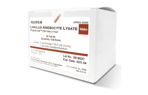
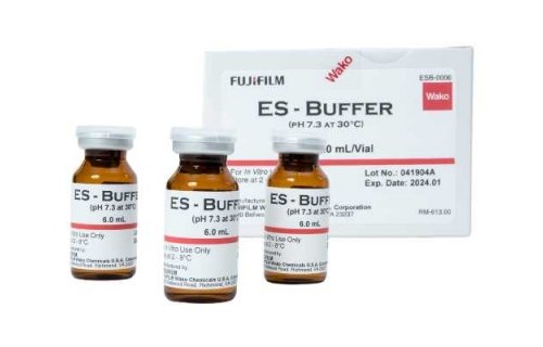
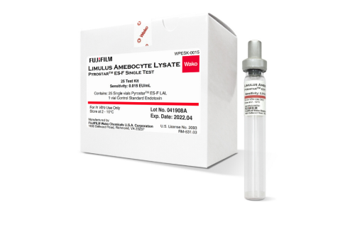
Additionally, a pH value that deviates from the optimal range of 6 to 8 can lead to protein denaturation and inactivation, thereby increasing the likelihood of obtaining false negative results.PYROSTAR™ has created a novel line of ES-F reagents, specifically targeting endotoxins, with the added benefit of ES-F buffer already incorporated into the formulation. PYROSTAR™ ES-F reagents are specifically designed to avoid reactions with (1→3)-β-D-glucan, minimizing the possibility of false positive results. Furthermore, these kits incorporate an endotoxin specific buffer, which utilizes Tris to neutralize both acids and bases, effectively maintaining the pH of the reaction mixture within the ideal range of 6–8. This pH control minimizes the likelihood of false negative results.
References
1.Neun, B.W., et al., Detection of Beta-Glucan Contamination in Nanotechnology-Based Formulations. Molecules, 2020. 25(15).
2.Barton, C., et al., Beta-glucan contamination of pharmaceutical products: How much should we accept? Cancer Immunol Immunother, 2016. 65(11): p. 1289-1301.
3.Roslansky, P.F. and T.J. Novitsky, Sensitivity of Limulus amebocyte lysate (LAL) to LAL-reactive glucans. J Clin Microbiol, 1991. 29(11): p. 2477-83.
4.Tran, T. and S.G. Beal, Application of the 1,3-β-D-Glucan (Fungitell) Assay in the Diagnosis of Invasive Fungal Infections. Arch Pathol Lab Med, 2016. 140(2): p. 181-5.
About the author:
 Lisa Komski, General Manager LAL sales
Lisa Komski is the Sales General Manager for the LAL Division of FUJIFILM Wako Chemicals U.S.A. Corporation. With a nearly 30-year career of working in the Chemicals and Life Science industries, she has established herself as a strong business development professional with a keen focus on customer service. As a child, Lisa dreamed of becoming a doctor, so having the opportunity to manage the PYROSTARTM line for the past 6 years has allowed her to be involved in a dynamic business that supports her lifelong passion to help protect the health and safety of others. Lisa holds degrees in Biology and Medical Technology and is also fluent in both the English and Spanish languages. When having the opportunity to take some personal time, Lisa enjoys family, friends, travel abroad and spoiling her adorable grandson. Contact Me - LinkedIn Profile