Common lab sources of Endotoxin and strategies for its detection and removal
Bacterial endotoxins, which are strong pyrogens ubiquitously present in the environment, also represent a major cause of concern as lab contaminants. If endotoxins remain undetected after contaminating lab samples, materials, or equipment, they will lead to unreliable and misleading experimental results. Moreover, if injectable products become contaminated with endotoxin, they can cause a severe pyrogenic response in the recipients.1
How does endotoxin contamination arise in the laboratory?
Bacterial endotoxins are constituents of the outer membrane of gram-negative bacteria and are shedded in small amounts during the life cycle of the bacteria and in large amounts after their death. It has been estimated that a single E. coli bacterium may contain up to 2 million endotoxin molecules.2 Endotoxins may contaminate laboratories via different routes, including contaminated water, air, or human skin.3 Endotoxin contamination may spread to lab plasticware or glassware as well as to widely used lab reagents and solutions, including cell culture media, fetal bovine serum, recombinant growth factors, and dissociation reagents.
Why is endotoxin contamination in the lab dangerous?
Endotoxin contamination of cell cultures or experimental samples would lead to variable and misleading experimental results. The pyrogenic activity of endotoxins can mask the inherent properties of cells by stimulating the release of tissue factors. Moreover, endotoxins elicit an inflammatory and pyrogenic response in vivo. This is why endotoxin contamination of injectable products may provoke a severe and potentially even life-threatening reaction in the recipients.
Endotoxin contamination testing in lab settings
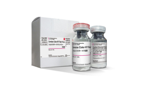
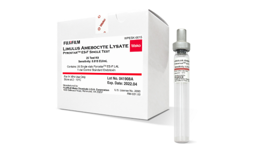
Several different methods for endotoxin detection exist, among which the Limulus amebocyte lysate (LAL) assay is most widely established. It relies on incubation of an endotoxin-containing sample with protein extracted from horseshoe crab blood, leading to a clotting reaction that indicates the presence of endotoxin.4 Depending on the LAL assay parameters, the endotoxin detection may be qualitative or quantitative, whereas its visualization may be turbidimetric or colorimetric.
Strategies for the removal of endotoxin contamination
Endotoxins are heat-resistant, which renders their removal difficult. Preventive measures, including the use of depyrogenated glassware, nonpyrogenic plasticware, and high-purity water for lab experiments, would help to avoid endotoxin contamination and to ensure the acquisition of reliable experimental data.
Discarding endotoxin-contaminated materials
The removal of endotoxin contamination is difficult, and if residual contamination remains, it may have serious consequences. Thus, if endotoxin-contaminated materials can be readily replaced with new ones, it would be prudent to discard them.
Decontamination of endotoxin-contaminated materials
Due to the heat resistance of endotoxins, standard autoclavation or sterilization protocols would be insufficient for endotoxin removal. However, dry heat exposure at higher temperatures and during a longer time period (such as 250°C for 30 min or at 180°C for 4 h) has been successfully used for endotoxin removal.4,5 Other protocols for endotoxin decontamination include the use of affinity chromatography and Triton X-114–based solutions.6,7 As alternative approaches, a cycle of washes in NaOH, HCl, and 70% ethanol or washes in 70% ethanol, which may be followed by a wash in acetic acid, have been proposed.3
Literature sources
- Li Y, Boraschi D. Endotoxin contamination: a key element in the interpretation of nanosafety studies. Nanomedicine (Lond). 2016;11(3):269-87. doi: 10.2217/nnm.15.196. Erratum in: Nanomedicine (Lond). 2016;11(6):739.
- Rhee SH. Lipopolysaccharide: basic biochemistry, intracellular signaling, and physiological impacts in the gut. Intest Res. 2014;12(2):90-5. doi: 10.5217/ir.2014.12.2.90.
- Gorbet MB, Sefton MV. Endotoxin: The uninvited guest. Biomaterials. 2005;26(34):6811–6817. doi: 10.1016/j.biomaterials.2005.04.063.
- https://www.fda.gov/inspections-compliance-enforcement-and-criminal-investigations/inspection-technical-guides/bacterial-endotoxinspyrogens.
- Sandle T. A comparative study of different methods for endotoxin destruction. Am Pharm Rev. 2013;Supplement.
- Schneier M, Razdan S, Miller AM, Briceno ME, Barua S. Current technologies to endotoxin detection and removal for biopharmaceutical purification. Biotechnol Bioeng. 2020;117(8):2588-2609. doi: 10.1002/bit.27362.
- Teodorowicz M, Perdijk O, Verhoek I, Govers C, Savelkoul HF, Tang Y, Wichers H, Broersen K. Optimized Triton X-114 assisted lipopolysaccharide (LPS) removal method reveals the immunomodulatory effect of food proteins. PLoS One. 2017;12(3):e0173778. doi: 10.1371/journal.pone.0173778