Top 5 tips for reducing Endotoxin Contamination in the lab
Cell culture systems can be contaminated by endotoxins, which are chemical contaminants produced by gram-negative bacteria and contain lipopolysaccharides. The presence of endotoxins is common in water, sera, and certain culture additives, particularly those produced using microbial fermentation. These toxins can be accurately measured using the Limulus Amoebocyte Lysate assay (LAL).[1]
Sources of endotoxins
Nonsterile Supplies, Media and Solutions: The unintended use of nonsterile supplies, media, or solutions during regular cell culture procedures is a primary cause of biological contaminants. Water, cell culture medium and reagents, serum, and plastic glassware are the primary sources of endotoxin contamination in the laboratory.[1]
Effect of endotoxin contamination
In vivo, these molecules with high biological reactivity have significant effects on both humoral and cellular systems. Using in vitro systems to study endotoxins has revealed their potential to influence the growth and performance of cultures, introducing a notable source of variability in experiments.[1]
Contamination with endotoxins can cause inaccurate measurements of gene expression or cellular function in the analysed samples. This occurrence has been noted in various cell types and materials, primarily because of the pyrogenic effects of endotoxins. One instance is when nanomaterials become contaminated with endotoxins, which can cause them to have inflammatory properties that interfere with accurately assessing their true biological properties.[2]
Tips for Minimizing Endotoxin Contamination
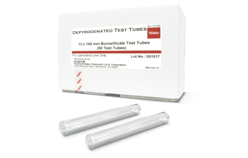
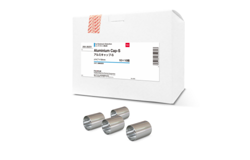
Depyrogenation of Glass Components
Vials and glass components have traditionally been made pyrogen-free through the process of dry heat sterilization at elevated temperatures. The depyrogenation of glassware and equipment is advised to be carried out by subjecting them to a temperature of 250°C for a duration of 45 minutes. Also, the destruction of pyrogens can be achieved by subjecting them to temperatures of 650°C for 1 minute or 180°C for 4 hours.[3]
Depyrogenation for Tubing and Plasticware
To eliminate pyrogens from plastic tubing, stir bars, and other tools that are not heat-resistant, it is recommended to flush them with Cavicide and then rinse them thoroughly with sterile water that is free from endotoxins.
To effectively eliminate any endotoxin residue adhering to the internal surfaces of the instrument, it is advantageous to run acetonitrile through an HPLC system subsequent to the use of sterile, depyrogenated water.[4]
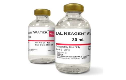
Water Purification
Distillation and Reverse Osmosis are the two approved methods of manufacturing for Water for Injection systems. It has been demonstrated that distillation is an effective and highly dependable technique for eliminating endotoxin from contaminated water samples.[3]
Endotoxin free reagents and media
To mitigate the possibility of endotoxin contamination, one should employ sera, reagents, and media that have been tested for endotoxins. Commercially developed sera, media, and reagents have been designed to prevent contamination by endotoxins, and they are tested for this purpose.[4]
Maintain Sterility
It is important to avoid introducing breath, coughs, or sneezes into the tubes or reaction beakers. It is also recommended to practice not touching your face. Despite the seemingly trivial nature of these precautions, they frequently contribute to contamination.[3, 5]
The presence of endotoxin contamination can result in inaccurate and unreliable experimental outcomes, impacting both diagnostic and research laboratories. By implementing straightforward measures such as endotoxin testing and utilizing non-pyrogenic plasticware, depyrogenated glassware, high-purity water, and endotoxin-tested cell culture reagents and media, one can effectively prevent endotoxin contamination and ensure the reliability of the acquired outcomes.
References
- Ryan, J.A., Understanding and managing cell culture contamination. 1994: Corning Incorporated.
- Li, Y. and D. Boraschi, Endotoxin Contamination: A Key Element in the Interpretation of Nanosafety Studies. Nanomedicine, 2016. 11(3): p. 269-287.
- Dept. of Health, E., and Welfare Public Health Service Food and Drug Administration, Bacterial Endotoxins/Pyrogens. FDA, 2014.
- (US), N.C.I., Endotoxin & Depyrogenation Tips. National Cancer Institute’s Nanotechnology Characterization Laboratory Assay Cascade Protocols, 2023.
- Schneier, M., et al., Current technologies to endotoxin detection and removal for biopharmaceutical purification. 2020. 117(8): p. 2588-2609.