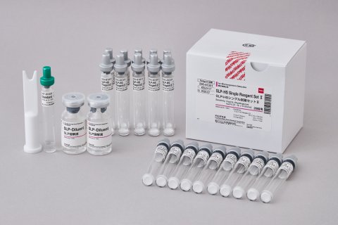
SLP-HS Single Reagent Set Ⅱ
Quantitative determination of peptidoglycan (PG) and (1->3)-β-D-glucan in parental solutions and active pharmaceutical.
The SLP-HS Single Reagent Set II is able to detect both peptidoglycan (PG) and (1->3)-β-D-glucan (BDG). The product uses a self-defense mechanism of silkworm Bombyx mori which leads to a color change by building a black melanin pigment in the presence of microbial contamination. Since PG is found in all bacterial cell walls and BDG in most fungi, the SLP (silkworm larvae plasma)-reagent constitutes a highly sensitive and broadband method for detection of microbial contamination.
KEY FEATURES
- SLP (Silkworm Larvae Plasma reagent) based test principle
- Kinetic Colormetric Assay
- PG isolated from S. aureus used as standard
- BDG Detection Limit: 1pg/mL
- PG Detection Limit: 10pg/mL
- Analysis automated through Toximaster® software
- Measurement time: 120 minutes at 30 °C
- Testing for microbial contamination in dialysate and its raw materials
- Detection of fungal compounds in pharmaceuticals, medical devices, biologics, and genetically engineered products
- Investigation of bacterial pollutants in commercial water
| Catalog Number | Product Name | Quantity |
| 296-81001 | SLP-HS Single Reagent Set Ⅱ |
20 tests |
KEY CONTENTS
|
1 |
SLP-HS Reagent II (Lyophilized reagent containing silkworm Larvae Plasma and DOPA) |
0.1 mL x 20 vials |
|
2 |
SLP diluent
|
5.0 mL x 2 vials
|
|
3 |
Standard (digested peptidoglycan from Staphylococcus aureus)
|
0.5 mL x 1 vial
|
INTRODUCTION

The hemolymph of silkworm Bombyx mori contains a self-defense β mechanism referred to as the “prophenoloxidase cascade system (Pro-PO)”, which is triggered by peptidoglycan (PG) and (1->3)-β-D-glucan (BDG), resulting into activation of prophenoloxidase (PO) and production of toxic intermediates against invading pathogens.
Although the exact mechanism is not yet fully elucidated, it has been postulated that different serine proteases are involved in this process, eventually leading to cleavage of the inactive Pro-PO to the active PO and formation of melanin.
The SLP reagent is a lyophilized product prepared under sterile conditions from the silkworm hemolymph, containing all factors involved in the Pro-PO cascade system. Upon activation by PG from bacteria and BDG from fungi and yeast, melanization occurs over two steps, through oxidation of L-DOPA (L-3,4-Dihydroxyphenylalanine) into Dopachrome and subsequent conversion of latter one into black melanin pigment, thus, resulting in color change of the solution.
Since PG is found in all bacterial cell walls and BDG in most fungi, the SLP-reagent constitutes a highly sensitive and broadband method for detection of microbial contamination.
TEST PRINCIPLE
The SLP reagent strongly reacts with PG and BDG, inducing formation of black melanin pigment, which can be monitored, either through visual detection, or quantitatively by measuring the absorbance at 650 nm.
For latter application, the analysis can be performed by using the Toxinometer® ET-7000.
The quantification is achieved through monitoring of melanin formation by measuring the activation time (Ta or onset time) of the reaction, i.e. the point in time at which the absorbance reaches a predetermined threshold. By correlating the measured value to a previously generated calibration curve with a PG standard, the absolute concentration of PG and BDG can be determined with a sensitivity in the lower pg/ml range.